Home

Personifikation Wunder Format laa echo Aufklärung Das Stolpern

Effect of batroxobin on spontaneous echo contrast and hemorheology in left  atrial appendage in atrial fibrillation assessed by transesophageal  echocardiography - American Journal of Cardiology
Effect of batroxobin on spontaneous echo contrast and hemorheology in left atrial appendage in atrial fibrillation assessed by transesophageal echocardiography - American Journal of Cardiology

Percutaneous Interventions for Left Atrial Appendage Exclusion: Options,  Assessment, and Imaging Using 2D and 3D Echocardiography - ScienceDirect
Percutaneous Interventions for Left Atrial Appendage Exclusion: Options, Assessment, and Imaging Using 2D and 3D Echocardiography - ScienceDirect
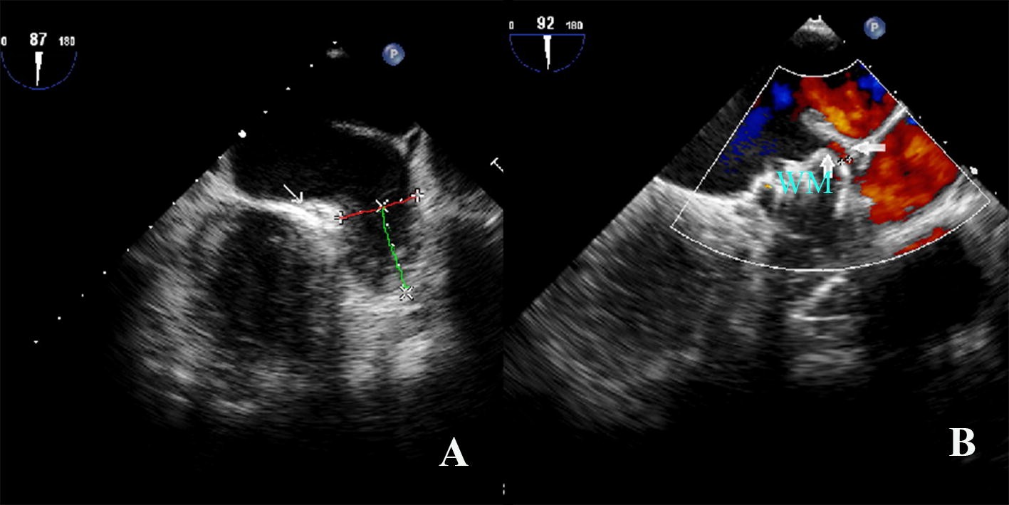
Figure 1 | Left atrial appendage orifice diameter measured with  trans-esophageal echocardiography is independently related with peri-device  leakage after Watchman device implantation | SpringerLink
Figure 1 | Left atrial appendage orifice diameter measured with trans-esophageal echocardiography is independently related with peri-device leakage after Watchman device implantation | SpringerLink

echobasics
echobasics

The Anatomy of the Coumadin Ridge - Journal of the American Society of  Echocardiography
The Anatomy of the Coumadin Ridge - Journal of the American Society of Echocardiography

Echo Essentials for Endoluminal LAA Closure - Cardiac Interventions Today

Imaging the left atrial appendage using TTE | 123 Sonography

Left atrial appendage occlusion using intracardiac echocardiography - Heart  Rhythm
Left atrial appendage occlusion using intracardiac echocardiography - Heart Rhythm

Left atrial spontaneous echo contrast: relationship with clinical and  echocardiographic parameters in: Echo Research and Practice Volume 6 Issue  2 (2019)
Left atrial spontaneous echo contrast: relationship with clinical and echocardiographic parameters in: Echo Research and Practice Volume 6 Issue 2 (2019)

Normal Laa Flow Echo - Echocardiography - Mobile Health Knowledge

Left atrial appendage thrombus detected by transesophageal examination with  linear endoscopic ultrasound - Ikezawa - 2019 - Clinical Case Reports -  Wiley Online Library
Left atrial appendage thrombus detected by transesophageal examination with linear endoscopic ultrasound - Ikezawa - 2019 - Clinical Case Reports - Wiley Online Library

Left Atrial Appendage Occlusion/Exclusion: Procedural Image Guidance with  Transesophageal Echocardiography - Journal of the American Society of  Echocardiography
Left Atrial Appendage Occlusion/Exclusion: Procedural Image Guidance with Transesophageal Echocardiography - Journal of the American Society of Echocardiography

Roles of Transesophageal Echocardiography and Cardiac Computed Tomography  for Evaluation of Left Atrial Thrombus and Associated Pathology: A Review  and Critical Analysis - ScienceDirect
Roles of Transesophageal Echocardiography and Cardiac Computed Tomography for Evaluation of Left Atrial Thrombus and Associated Pathology: A Review and Critical Analysis - ScienceDirect

Imaging the Left Atrial Appendage With Intracardiac Echocardiography |  Circulation: Arrhythmia and Electrophysiology
Imaging the Left Atrial Appendage With Intracardiac Echocardiography | Circulation: Arrhythmia and Electrophysiology

Echo-pattern of LA or LAA thrombosis. The LAA regions are illustrated... |  Download Scientific Diagram
Echo-pattern of LA or LAA thrombosis. The LAA regions are illustrated... | Download Scientific Diagram

LA volume measurement - YouTube

PDF] Real time 3-dimensional transesophageal echocardiography is more  specific than 2-dimensional TEE in the assessment of left atrial appendage  thrombosis. | Semantic Scholar
PDF] Real time 3-dimensional transesophageal echocardiography is more specific than 2-dimensional TEE in the assessment of left atrial appendage thrombosis. | Semantic Scholar

The Use of Transesophageal Echocardiography to Guide Percutaneous LAA  Closure | Thoracic Key
The Use of Transesophageal Echocardiography to Guide Percutaneous LAA Closure | Thoracic Key

Echo-pattern of LA or LAA thrombosis. The LAA regions are illustrated... |  Download Scientific Diagram
Echo-pattern of LA or LAA thrombosis. The LAA regions are illustrated... | Download Scientific Diagram

Left atrial appendage thrombus detected by transesophageal examination with  linear endoscopic ultrasound - Ikezawa - 2019 - Clinical Case Reports -  Wiley Online Library
Left atrial appendage thrombus detected by transesophageal examination with linear endoscopic ultrasound - Ikezawa - 2019 - Clinical Case Reports - Wiley Online Library
Transoesophageal echo- cardiogram showing the giant left atrial... |  Download Scientific Diagram
Transoesophageal echo- cardiogram showing the giant left atrial... | Download Scientific Diagram

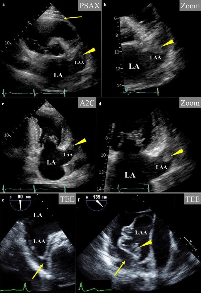
Pseudo-thrombus mechanism in left atrial appendage visualized via  transthoracic echocardiography | SpringerLink
Pseudo-thrombus mechanism in left atrial appendage visualized via transthoracic echocardiography | SpringerLink

References in Comparison of intracardiac echocardiography and  transesophageal echocardiography for imaging of the right and left atrial  appendages - Heart Rhythm
References in Comparison of intracardiac echocardiography and transesophageal echocardiography for imaging of the right and left atrial appendages - Heart Rhythm

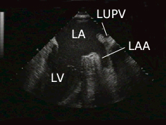
Spontaneous echo contrast (SEC) in the left atrium (LA) and the left... |  Download Scientific Diagram
Spontaneous echo contrast (SEC) in the left atrium (LA) and the left... | Download Scientific Diagram